Food Control: Research Progress Of Aptamer Sensor Technology In The Detection Of Foodborne Pathogenic Bacteria

Professor Wang Zhouping's team from the School of Food Science and Technology of Jiangnan University published a review paper titled "of in the of" (Research Progress of Aptamer Sensor Technology in the Detection of Foodborne Pathogenic Bacteria) in Food.

Summary:

In recent years, foodborne pathogenic bacteria have attracted increasing public concern. Therefore, constructing a simple, fast, effective, and low-cost rapid detection method has become an important issue in the field of food safety. Among them, aptasensors are emerging biosensors in the fields of food safety, detection and control. Nucleic acid aptamers (Nucleic acid aptamers) are single-stranded DNA or RNA sequences that specifically bind to proteins or other small molecules through systematic evolution of exponentially enriched ligands (SELEX) and other derivative technologies. Nucleic acid aptamers are widely used in food safety testing due to their wide range of target molecules, high sensitivity, strong specificity, and good stability. This article will review the latest research on aptamers of foodborne pathogenic bacteria, potential applications and optimization methods of aptasensors in foodborne pathogenic bacteria. Finally, we hope that this review can provide new ideas and approaches for the development and application of aptamer sensors for foodborne pathogenic bacteria.
Research on fit of tops_Research on fit of tops_Research on fit of tops

Fig. 1. of paper.

2.Adapter

Fig. 2. , and of the . (A) of the . (B) . (C) the of the L3 by a , with from Ref (Ma et al., 2022). (D) of the by a 2′- O-RNA base, from Ref (Maio et al., 2017).

4. Combination of aptamers and biosensors

Research on fit of tops_Research on fit of tops_Research on fit of tops

4.1 Application of aptamers in colorimetric sensors

Fig. 3. The -based . (A) based on label-free AuNPs (a), (b). (B) of for S., with from Ref (Wei et al., 2022). (C) n of the for of S. , from Ref (Yu et al., 2020).

4.2. Application of aptamers in fluorescence sensors
Research on fit of tops_Research on fit of tops_Research on fit of tops

Fig. 4. The -based . (A) of the S. , with from Ref (Ren et al., 2019). (B) for V. , from Ref (Wu, 2019). (C) for E. , with from Ref (Yao et al., 2021).

4.3. Application of aptamers in electrochemical sensors

Fig. 5. The -based . (A) for of C. , with from Ref (Peng et al., 2019). (B) for B. , from Ref ( et al., 2019). (C) the of E. coli, with from Ref (Hua et al., 2018).

4.6 Zoom in

Research on fit of tops_Research on fit of tops_Research on fit of tops

Fig. 6. The of the -based , and -based .(A) of V. , from Ref (Wu et al., 2015). (B) of P. , with from Ref (Xie etal., 2022). (C) for the of E. coli, from Ref (Yang et al., 2020).

5. Conclusion Future Prospects and Challenges

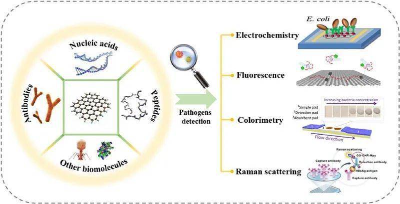
Foodborne pathogenic bacteria are an important threat to food safety. In summary, this article reviews the research progress of aptamers in food-borne pathogenic bacteria in recent years, as well as the application of aptamer sensors in the detection of food-borne pathogenic bacteria. These aptamer-based biosensors exhibit good detection sensitivity, selectivity, and accuracy by introducing new signal transduction systems and multiple amplification strategies. We can find that colorimetric and fluorescent sensors have similar detection limits. But fluorescent sensors tend to have a larger detection range, while electrochemical sensors have the highest sensitivity and the lowest detection limit. However, during the application process, there are still some challenges that need to be solved in the application of these biosensors.

With the development of SELEX technology, the selection of faster and more accurate nucleic acid aptamers and a series of nucleic acid aptamer optimization methods are also conducive to obtaining more stable, better performance, higher affinity, and lower costs. of nucleic acid aptamers. Currently, in terms of aptamers, obtaining more stable and higher affinity aptamers is the direction of development. To achieve this goal, analyzing the specific binding sites of bacterial aptamers and the specific components of bacterial whole cells has become a key issue. However, due to the complexity of bacteria, there are few studies on the analysis of aptamer-bacterial binding sites and the molecular resolution of bacterial-aptamer binding. In order to solve the above problems, the application of computational prediction tools has become particularly important. In addition to using computers for primary sequence analysis, two-dimensional and three-dimensional structure prediction, how to complete the docking of complex targets such as food-borne pathogenic bacteria and aptamers and conduct corresponding molecular dynamics simulations to more accurately evaluate aptamers The stability of the body-target complex and determining its binding energy are also future considerations. Continuing developments along this direction could enhance the practical significance of aptamers.
Research on fit of tops_Research on fit of tops_Research on fit of tops

In terms of sensors, aptamer sensors targeting the detection of foodborne pathogenic bacteria have been a hot research topic in recent years. Optical aptamer sensors can detect changes in color or absorbance by direct observation. The electrochemical aptasensor has high sensitivity. A large number of papers have been published in this field, but most of them are verification of conceptual models and lack the potential for clinical practice. Therefore, it is necessary to improve aptasensor technology and sensor practicality. On the one hand, due to the complex composition of actual samples and the influence of environmental factors, the structure and properties of aptamers will change. Such as protein, lipid carbohydrates, pH and temperature, etc.

Nonspecific binding of aptamers may also lead to false-positive results, a phenomenon known as matrix effect. For example, target substances in milk can easily combine with other proteins to form polymers, thus affecting the detection results. This problem is often solved by diluting the sample before analysis or by protein precipitation. However, both processes are time-consuming. Therefore, it is necessary to study the effects of environmental factors, actual sample compositions of proteins and lipids on aptamer-target binding to guide the application of aptamers in detection systems. On the other hand, detected foodborne pathogens can present false positives due to mutations. Selecting aptamers that target specific components of bacteria can help avoid mutations. For example, bacteria can be identified through targeted screening of aptamers targeting specific proteins or polysaccharide components.

Sensors with high sensitivity are usually achieved by using one or more signal amplification methods. However, different amplification methods also have their limitations. Magnetic enrichment of magnetic species may affect signal intensity, and combination with enzymatic reactions may hinder the sensor's ability to function optimally in real-world and complex samples. The main challenge with nanomaterials is ensuring sensor stability and fabrication reproducibility. The solution to these problems is also the future development direction of fitness sensors.

Original link:

Online submission platform link:

Liu Juewen: New Research On A Classic Aptamer—binding Site, Cooperativity, And More Sensitive Adenosine

In 1995, scientists first discovered an adenosine aptamer with a length of 27 nt. So far, this aptamer has been widely used in biophysical research and the development of biosensors. NMR results show that the aptamer has two identical binding sites, while most small molecule aptamers only have one binding site. Adjusting the number of binding sites is of great significance in affecting the sensitivity of the biosensor. Therefore, Liu et al. hope to experimentally explore whether the two adenosine binding sites are independent and cooperative, and measure the thermodynamic parameters of the binding process through isothermal titration calorimetry ITC to improve the detection performance of the biosensor.

The secondary structure of the initially selected adenosine aptamer is shown in Figure 1A, named Apt2a. It can bind to two adenosine molecules, and the two binding sites are "Site1" and "Site2" respectively. According to its NMR results, each adenosine can interact with two nearby nucleotides through hydrogen bonds and overlap with guanine through reverse mismatching, and these two sites have the same binding effect. Subsequently, the research team used isothermal titration calorimetry ITC to characterize the thermodynamic properties of its binding process. Using this label-free technology, the Kd value of the wild-type aptamer was found to be 16.4 μM, and each aptamer was able to bind 2.1±0.2 adenosine molecules (Figure 2A).

Figure 1 Schematic diagram of secondary structures and binding sites of wild-type and mutant aptamers.

Figure 2 Relevant thermodynamic parameters and specific characterization of wild-type and mutant aptamers

After confirming the binding thermodynamic properties, the research team wanted to verify whether one of the binding sites could be removed while the binding affinity and specificity of the other site were still retained. Therefore, a G5T mutant was introduced in the study, in which a C base was inserted to pair with G22, resulting in an increase of two base pairs. As shown in Figure 1B, this mutant is called Apt1a, and it is speculated that the base adjustment in it will eliminate the presence of binding site 1. The ITC results showed that the Kd value of mutant Apt1a was 12.0 μM, and each aptamer was able to bind 1.1±0.1 adenosine molecules, which was in line with the expected design. Both wild-type and mutant aptamers bind to the target with specificity, and there is no thermal response when adding cytidine or guanosine (Figure 2A and B, red and blue lines). Therefore, the research team successfully eliminated adenosine binding site 1 and retained binding site 2, experimentally proving for the first time that the classic adenosine wild-type aptamer can be transformed into a single-site aptamer.

Subsequently, the research team experimentally verified the thermodynamic equivalence of the two binding sites. They used the same approach to design Apt1b, retaining adenosine binding site 1 and blocking binding site 2 (Figure 1C). The Kd value was determined to be 14.1 μM through ITC experiments, and each aptamer was able to bind 0.8±0.2 adenosine molecules, proving that the two binding sites have similar binding affinity. Although there are certain differences in the entropy and enthalpy of the binding reaction, the two binding sites are basically equivalent.

In the above experiments, the research team blocked the response binding site by introducing new base pairs and replacing base pairs, but the stable structure of the blocked binding site was still retained. Next, the research team tried to destroy the stable structure of its binding site by deleting specific base pairs, thereby removing the binding site. For example, they deleted the first three base pairs of the wild-type Apt2a aptamer, which also disrupted the target-binding ability of the Apt2b mutant (Figure 1D and Figure 2D). This experiment demonstrates that disrupting the structure of one junction site causes the second binding site to also be blocked, even though the second binding site is structurally intact. Therefore, for wild-type aptamers, any binding site can bind adenosine first, and the binding of any site can also stabilize the other site. Due to similar Kd values, it is unlikely that the two binding sites have a preference in binding order. In addition, the research team optimized the aptamer concentration and reaction temperature through ITC results. The optimal conditions for the reaction were 10 μM aptamer binding at 10°C (Figure 3).

Figure 3 Temperature optimization

In previous studies, this adenosine aptamer has been widely used in the development of biosensors. However, through the above studies, one binding site can be eliminated and shorter aptamers can be used for biosensing. For example, although the wild-type aptamer cannot be truncated, three base pairs can be deleted from the Apt1a mutant to generate Apt1c (Figure 1E), and ITC results indicate that the aptamer still retains the binding effect of the corresponding region (Figure 2E ), have similar Kd values ​​and bind only one adenosine. This mutant has only 21 nt compared to the 27 nt wild-type aptamer.

In addition, as shown in Figure 4, by directly fitting the above ITC data, it was found that the Hill coefficients of the three mutants were approximately 1, while that of the wild type was approximately 1.2. Therefore, it is shown that the two binding sites have a very weak cooperative relationship. In addition, the structures of Apt3 and Apt4 mutants are shown in Figure 1F-G. The Hill coefficients of the two are close to 2. It is speculated that the proximity of the two sets of binding sites may lead to enhanced cooperativity.

Figure 4 Relevant thermodynamic parameters of wild-type and mutant aptamers

Biosensors built using multi-binding site aptamers have low sensitivity when analyzing low concentrations. Therefore, this study is the first attempt to use single-binding site adenosine aptamers for biosensors to build a more sensitive biosensor by reducing the number of binding sites. As shown in Figure 5A, the Apt2a and Apt1a aptamer sequences were extended respectively, and the extended sequence was hybridized with a fluorescent group-labeled fragment (F-DNA). Part of the extended sequence and part of the aptamer sequence were hybridized with a quenching group-labeled fragment (F-DNA). Q-DNA) hybridization. In the presence of target adenosine, the nucleic acid aptamer folds and releases the quenching chain, resulting in enhanced fluorescence, thereby achieving quantitative detection.

Figure 5 Schematic diagram of biosensor principle

As shown in Figure 6A-B, the fluorescence intensity reached the maximum value 30 minutes after adding the target, and the adenosine concentration-maximum fluorescence intensity function graph drawn from the results is shown in Figure 6C. In the presence of low concentrations of adenosine, there was a roughly linear relationship between target concentration and fluorescence intensity (Figure 6D). The slope of the curve of the Apt1a mutant was 3.8 times that of wild-type Apt2a, and both groups showed good target specificity (Figure 6E-F). The biosensor constructed based on mutant Apt1a has a detection limit of adenosine of 9.1 μM.

Figure 6 Biosensor performance characterization

In summary, this study used reasonable sequence design to remove each binding site of the adenosine aptamer individually, and verified that each group has similar binding affinity and specificity through isothermal titration calorimetry ITC, and discussed its biochemical significance. Additionally, the number of target binding sites can be increased, with up to four sites introduced in a single DNA sequence. Moreover, different aptamer sequences can also be used to assemble fluorescent biosensors. At lower adenosine concentrations, the sensitivity of the single-site aptamer increased 3.8-fold, with a detection limit of 9.1 μM adenosine. This work provides a solution for studying the relationship between the number of aptamer binding sites and detection sensitivity, and also has great reference significance for the field of biosensors.

Comments:

1. This article modified the classic adenosine aptamer, explored the independence and cooperation between different binding sites of the aptamer, and realized the optimization and tailoring of the aptamer, which is of great innovative significance;

2. Through reasonable sequence design, this paper uses the modified aptamer to construct a fluorescent biosensor. At a lower adenosine concentration, the sensitivity of the single-site aptamer is increased by 3.8 times, and the detection limit is 9.1 μM;

3. This article has innovative concepts, clear ideas, complete characterization, and combines thermodynamics with aptamer research to enrich related characterization methods.